96 Hour Chick Embryo Serial Section
воскресенье 24 февраля admin 54
In the stage from 22 hours on, the somites formed in the mesoderm at the left and right side of the neural walls become visible. Active disk image key. After 24 hours 4 to 5 segmented paired blocks can be discerned. Later on, these structures will differentiate into the vertebrae, the ribs, a part of the skin and the dorsal muscles. Only the head region lifts up above the area pellucida. In this preparation, one can see the chorda (notochord) in the region of the anterior intestinal portal.
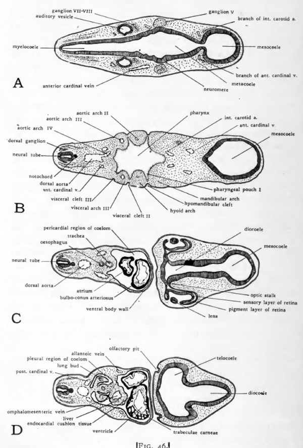
Of chick embryos were analyzed by study of serial cross-sections of a continuous series of normal embryos between 40 to 72 hours of incubation. Two extirpation experi- ments were. The neural tube, at 48, 72, 96 or 120 hours of incubation. Serial sections are where we slice an embryo as if it was a sausage. We affix each section, in order, on a microscope slide and stain the sections before we look at them with the microscope. We then 'read' these two-dimensional sections to try to visualize the three-dimensional organization of all the layers and parts of an embryo at one stage.
This structure marks the differentiating foregut which is formed as a blind pocket bordered by endodermal tissue. The neural walls end in a neural pore at the anterior side and become smaller and wider apart in the region of Hensen’s node where it ends in the sinus rhomboidalis. Sometimes the extra-embryonic vessels become already visible in the area vasculosa. Later on, they will make contact with the vitelline (omphalomesenteric) veins and arteries formed in the embryo.
• Developmental stages after 22-28 hrs, according to Patten (1920) • Whole mount preparation 24 hours () • Cross sections 24 hours () Developemental stages 22-28 hrs according to Patten (1920) Dorsal view of a developing chicken embryo (between 22 - 28 hrs after fertilization) • 22 to 23 hrs: the beginning of somite formation • 24 hrs: 4 pairs of mesodermic somites are visible • 27-28 hrs: 8 pairs of mesodermic somites are visible Stage 24 hours Whole mount preparation 24 hours Information: The somites are formed in the mesoderm at the left and right side of the neural walls. In this stage, they are visible as 4 to 5 segmented paired blocks. Afterwards these structures will differentiate in to the vertebrae, the ribs, a part of the skin and the dorsal muscles. Only this head region elevates above the underlying area pellucida.
In this preparation, one can see the chorda (notochord) in the region of the differentiating foregut. Embryology of the chicken 24 hours after fertilization Right: stained whole mount preparation. Herebelow A and B: cross sections at the level of the primitieve groove and the neural groove.
At about 33 hours after fertilization, the embryo is about 4 mm long and the first flexion of the originally straight embryo starts in the head region. The cranial flexure will be visible a few hours later. At this stage 12 to 13 somites are formed. The eye vesicles are rather large. The forebrain vesicle or prosencephalon will divide, the midbrain vesicle or mesencephalon remains undivided while the hindbrain vesicle or rhombencephalon will form a series of smaller neuromeres.
The sinus rhomboidalis (diamond-shaped) is still present as the only opening of the neural tube and the primitive streak is only rudimentary. The infundibulum (= derived from the diencephalon) appears as a half circular structure at the ventral side of the caudal part of the forebrain. The notochord or chorda dorsalis ends just behind this ventral vesicle. Later on, 36 hours after fertilization, the heart, which has a bilateral origin in the mesodermal layer, is a S-shaped tube which protrudes to the right of the embryo (in upper view).
Outside, in the area vasculosa (= forseen of blood vessels) the formation of blood islands continues. The primitive streak can only still be discerned below the sinus rhomboidalis. Early embryonic developmental features that are shown on this page: • Whole mount preparation 33 hours () • Whole mount preparation 36 hours () • Cross sections 36 hrs; formation of eye, heart and intestines ( ) Stage 33 hours Information: At about 33 hours after fertilization, the embryo is about 4 mm long and the first flexion of the originally straight embryo starts in the head region and the cranial flexure will be visible a few hours later. At this stage 12 to 13 somites are formed.
The eye vesicles are rather large. The forebrain vesicle or prosencephalon will divide, the midbrain vesicle or mesencephalon remains undivided while the hindbrain vesicle or rhombencephalon will form a series of smaller neuromeres. The sinus rhomboidalis (diamond-shaped???) is still present as the only opening of the neural tube and the primitive streak is only rudimentary.
The infundibulum (= derived from the diencephalon) appears as a half circular structure at the ventral side of caudal part of the forebrain. The notochord or chorda dorsalis ends just behind this venral vesicle. Embryology of chicken 33 hours after fertilization: stained whole mout preparation 1 = Proamnion, 2 = Prosencephalon, 3 = Mesencephalon, 4 = Rhombencephalon, 5 = Somite, 6 = Eye vesicle, 7 = Foregut, 8 = Chorda (translucent), 9 = Heart, 10 = Lateral mesoderm, 11 = Spine, 12 = Sinus rhomboidalis, 13 = Primitive streakp, 14 = Blood islands Dorsalview and longitudinalsection at 33 hrs according to Patten Patten, B.M. The Early Embryology of the Chick. Philadelphia: P. Blakiston's Son and Co. Stage 36 hours Information: 36-hours after fertilization, the heart is a S-shaped tube which protude to the right of the embryo (in upper view).